
A new way to see fresh tissue.
PolaSight provides a direct, label-free visualization of fresh tissue, revealing physical contrast beyond what is preserved in a conventional stained slide.




H&E shows morphology.
PolaSight adds quantitative contrast.
Today's pathology is read from a stained glass slide. PolaSight reveals the fresh tissue's physical contrast directly and label-free - a complementary layer, not a stain.
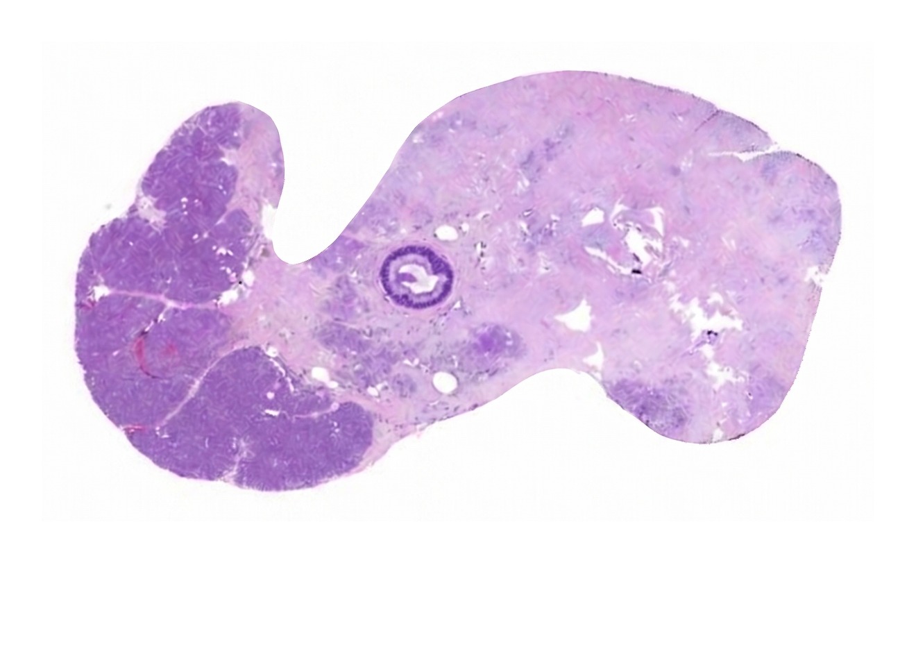
Morphology, fixed onto glass and read by eye - the standard stained section for over a century.
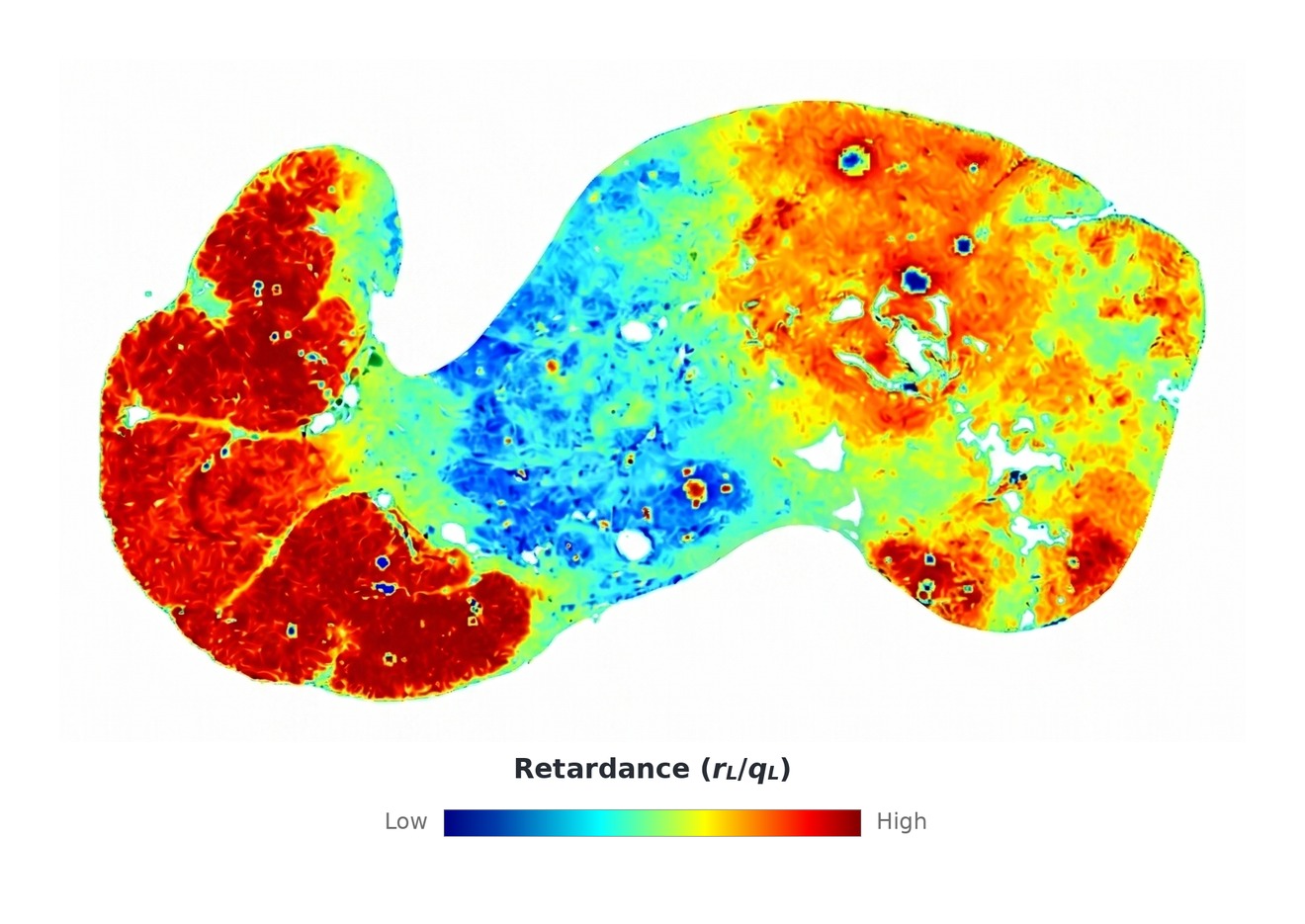
A quantitative, label-free signal from fresh tissue - data, not a reinterpretation.
The same tissue, two signals - H&E morphology and a label-free Mueller-matrix polarimetry (MMP) map (rL/qL retardance).
A new, structured layer of tissue data.
Every scan produces a quantitative, standardized record of fresh tissue - mapping optical properties across the sample, captured at the point of care.
Quantitative
Measuring optical properties such as retardance, depolarization, and scattering across the tissue.
Reproducible
The same tissue gives the same numbers - lab to lab, reader to reader.
AI-ready
Consistent, structured inputs - ready for computational analysis.
The platform that captures it.
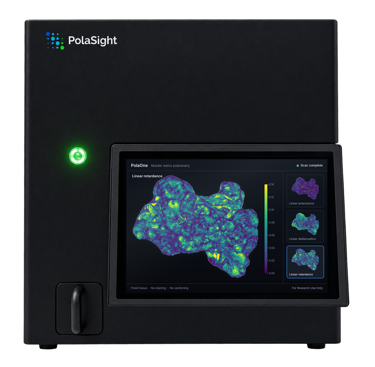
Label-free, polarized-light imaging of fresh tissue, right at the bench.
Place
Fresh tissue in a standard cassette. No staining, freezing, or slides.
Scan
One button. A fully automated polarimetric acquisition in about a minute.
View
Label-free maps of tissue microstructure, viewed alongside the slide.
Non-destructive - the specimen is preserved for downstream H&E and molecular work. For research use only.
First focus: fresh frozen-section tissue.
Our first research focus is fresh tissue at frozen section, imaged label-free without freezing or staining. The same imaging approach extends to other fresh-tissue research settings.
Multi-spectral polarimetric imaging.
PolaOne uses Mueller-matrix polarimetry, an established, label-free optical method that captures polarization-based contrast related to tissue microstructure and has been studied in peer-reviewed biomedical optics research for decades.
Label-free and reagent-free
No stains, no consumables, no tissue preparation - a direct optical view of the tissue itself, preserved intact for everything downstream.
Grounded in peer-reviewed science.
Built on peer-reviewed foundational research and an exclusive University of Bern license, with multi-center research studies now underway. Publication references available on request.
For research use only
PolaOne is an investigational device for research use only. Not for diagnostic or clinical use.
Pathology is going digital.
For over a century, tissue has been read from a single stained image. Fresh tissue holds a quantitative signal no one has captured - a physical layer beyond the stain. PolaSight captures it.
Built by pathology, optics, and AI.
A founding team pairing medtech operating experience with University of Bern science.




Supported by a medical advisory team at the University of Bern, including Prof. Aurel Perren (pathology) and Prof. Daniel Candinas (surgery).
Explore the imaging with us.
Tell us about your lab, your study, or your thesis - we read every message.
- Pathology & laboratory partners
- Research collaborators
- Investors